2 The Anti-Xa Assay
A five-minute revision chapter for FRCPath and FCPS
anti-Xa, UFH, LMWH, DOAC, haemostasis, FRCPath, FCPS
2.1 Learning objectives
By the end of this chapter, you should be able to:
- explain the chromogenic principle of an anti-Xa assay;
- select the correct indication, calibrator and sampling time;
- interpret UFH, LMWH and direct Xa-inhibitor results safely;
- recognise analytical and pre-analytical traps; and
- describe the next clinical or laboratory step after an unexpected result.
Anti-Xa assays measure inhibition of factor Xa, but the number is meaningful only when the drug, calibrator, sample timing and local decision range are known.
2.2 1. Test principle
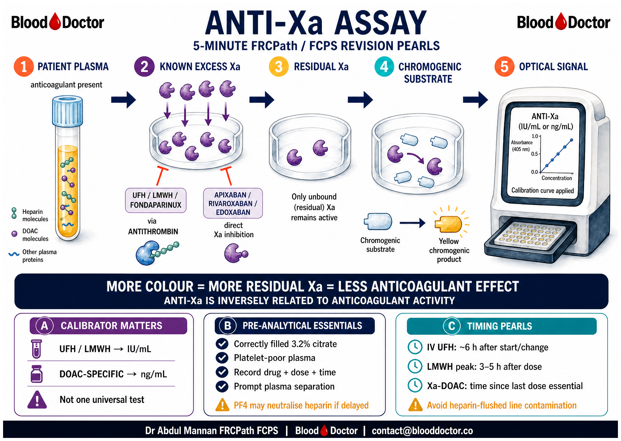
The anti-Xa assay is a chromogenic functional assay, not a clotting-time assay. A known excess of factor Xa is added to the patient’s citrated plasma. Anticoagulant present in the sample inhibits a proportion of that Xa. Residual, uninhibited Xa cleaves a chromogenic substrate and produces colour, which the analyser measures photometrically.
More colour → more residual Xa → less anticoagulant effect.
Less colour → less residual Xa → more anticoagulant effect.
The measured optical signal is converted to a reported result by a calibration curve. That curve determines the meaning and unit of the result:
| Assay calibration | What it measures | Typical report unit |
|---|---|---|
| UFH or LMWH | Heparin-mediated Xa inhibition | IU/mL |
| Fondaparinux | Fondaparinux effect | Drug-specific unit or concentration, according to local method |
| Apixaban, rivaroxaban or edoxaban | Direct Xa-inhibitor concentration | ng/mL |
2.2.1 Mechanistic distinction
- UFH, LMWH and fondaparinux potentiate antithrombin-mediated inhibition of Xa. UFH also has appreciable anti-IIa activity because sufficiently long heparin chains can bridge antithrombin and thrombin.
- Apixaban, rivaroxaban and edoxaban inhibit Xa directly and do not require antithrombin.
- Some anti-Xa methods add exogenous antithrombin. This affects how antithrombin deficiency influences the result, so know your local reagent design before drawing conclusions.
Say: “The assay measures residual factor Xa activity by chromogenic substrate cleavage, so absorbance is inversely related to anticoagulant effect.” This is clearer than saying only that the test “measures heparin level.”
2.3 2. Indications
2.3.1 UFH: when anti-Xa is particularly useful
Use anti-Xa to monitor a therapeutic UFH infusion when APTT-based monitoring is unreliable, for example with:
- a lupus anticoagulant or a pre-existing prolonged APTT;
- a raised factor VIII level in acute inflammation, which may shorten the APTT despite heparin effect;
- a factor deficiency, DIC, or another condition that confounds the APTT; or
- discordance between dose, APTT and the clinical picture, including suspected apparent heparin resistance.
2.3.2 LMWH: selective, not routine
Routine LMWH monitoring is not indicated in most stable adults. A properly timed level may be helpful in selected patients, such as those with severe renal impairment, pregnancy, extremes of body weight, paediatric practice, recurrent thrombosis or unexpected bleeding while treated.
2.3.3 Direct Xa inhibitors: answer a specific urgent question
A drug-specific calibrated anti-Xa assay can quantify apixaban, rivaroxaban or edoxaban when the result may change management, for example:
- life-threatening bleeding;
- urgent surgery or an invasive procedure;
- possible thrombolysis where recent exposure is uncertain;
- suspected overdose, accumulation, malabsorption or non-adherence; or
- major renal or hepatic deterioration.
Do not order an anti-Xa assay to monitor a stable patient on a DOAC routinely, and do not expect a standard anti-Xa assay to assess dabigatran. Dabigatran is a direct thrombin inhibitor: use a thrombin-based method such as a dilute thrombin time or ecarin-based assay where available.
2.4 3. How to perform the test
2.4.1 Before sampling: establish the clinical question
The request should state the anticoagulant, dose, route, exact time of the last dose, sampling time, renal function and reason for testing. This information is part of the test, not administrative detail.
2.4.2 Sample and handling
- Obtain a correctly filled 3.2% sodium citrate sample.
- Avoid heparin-contaminated lines. If a line must be used, follow the local discard and flushing policy.
- Produce platelet-poor plasma using the laboratory’s validated centrifugation process.
- Process promptly. Delayed separation can expose heparin to platelet factor 4, which may neutralise heparin and produce a falsely low result.
2.4.3 Timing
| Treatment | Practical timing principle |
|---|---|
| IV UFH infusion | Usually assess about 6 hours after starting or changing the infusion, then follow the local nomogram |
| Therapeutic LMWH | If a peak is requested, sample about 3–5 hours after the dose, ideally at steady state |
| Xa-DOAC | Record time since last dose. A number without timing cannot distinguish expected peak from trough exposure |
“Apixaban 5 mg twice daily; last dose 08:00; sample 12:00; urgent laparotomy planned; please perform apixaban-calibrated anti-Xa if available.”
2.5 4. Interpretation
2.5.1 UFH
Many institutions use an anti-Xa target around 0.3–0.7 IU/mL for therapeutic UFH. Treat this only as a familiar working range: local assay validation and the institutional UFH nomogram govern dosing.
| UFH anti-Xa result | Interpret with | Immediate next step |
|---|---|---|
| Below local target | timing, infusion delivery, sample source, weight-based dosing and residual Xa-DOAC exposure | correct a sampling or delivery issue first; then adjust using the local nomogram |
| Within local target | bleeding/thrombotic state and timing | continue and monitor as locally specified |
| Above local target | bleeding, renal and hepatic function, dose history and possible contamination | hold or reduce infusion according to protocol; assess for bleeding; repeat as indicated |
2.5.2 LMWH
Interpret only against the specific preparation, dose regimen, indication, timing and local target. A peak level from twice-daily therapeutic enoxaparin cannot be interpreted using a once-daily target, and a trough must not be called “subtherapeutic” simply because it is lower than a peak range.
2.5.3 Xa-DOACs
A drug-specific anti-Xa result is a concentration, not an automatic instruction to increase or decrease the dose. There is no universal therapeutic range for routine dose titration. Interpret the result in relation to dose timing, expected population exposure, renal function, interaction risk and the urgent clinical decision.
“Anti-Xa 0.6 IU/mL” from a UFH-calibrated assay is not interchangeable with “apixaban 60 ng/mL” from an apixaban-calibrated assay.
2.6 5. Clinical applications: what to do after the result
2.6.1 Scenario A: low UFH anti-Xa
- Check that sampling occurred after the appropriate equilibration period.
- Check infusion pump, prescription, actual dose, line source and body-weight calculation.
- Ask explicitly about apixaban, rivaroxaban or edoxaban in the previous week. These usually raise, rather than lower, a heparin-calibrated anti-Xa, but the history changes the interpretation of any discordance.
- If the result is reliable, adjust UFH only through the local protocol. Consider antithrombin testing if reduced heparin response is clinically plausible.
2.6.2 Scenario B: high UFH anti-Xa
- Assess the patient for bleeding and haemodynamic instability.
- Confirm that the sample was not contaminated by a heparinised line.
- Review recent Xa-DOAC use. Residual drug can cause apparent excess “heparin” activity.
- Hold or reduce UFH according to local protocol. Major bleeding or urgent reversal requires immediate senior haemostasis input; protamine decisions are clinical, not assay-only decisions.
2.6.3 Scenario C: unexpected LMWH peak
- Verify that it was a true peak sample and that the correct LMWH calibration was used.
- Review renal function, dose, weight and clinical bleeding or thrombotic events.
- Repeat a correctly timed sample if interpretation remains uncertain.
- Make dose or interval changes only through the relevant local protocol or specialist team.
2.6.4 Scenario D: Xa-DOAC level before an urgent procedure
- Confirm the drug, last dose, renal function and procedure bleeding risk.
- Request the matching drug-specific assay if a quantitative answer is needed.
- Discuss the result with the procedure and haemostasis teams. The decision may be to delay, proceed, reverse or choose an alternative approach.
- Do not infer a precise drug concentration from a heparin-calibrated result unless that relationship has been locally validated.
2.7 6. Errors, interferences and troubleshooting
| Unexpected finding | Possible cause | Safe next action |
|---|---|---|
| Markedly high heparin anti-Xa from a line sample | Heparin contamination | repeat from a clean sample before changing dose, if clinically safe |
| Low heparin anti-Xa after delayed processing | PF4-mediated heparin neutralisation | repeat using promptly processed platelet-poor plasma |
| High UFH anti-Xa just after UFH starts | Residual apixaban/rivaroxaban/edoxaban | suspect DOAC interference; establish a baseline and use the local transition strategy |
| Result does not match the request | Wrong calibration curve | ask the laboratory which calibration was used; do not act on the number until clarified |
| Optical flag or implausible result | Haemolysis, icterus or lipaemia, depending on assay design | discuss with the laboratory and repeat or use a validated alternative if required |
| Low heparin effect despite high dose | Antithrombin deficiency, altered clearance, under-delivery or timing error | verify delivery and sample; consider antithrombin deficiency in the appropriate clinical setting |
| Undetectable anti-Xa but anticoagulant effect suspected | Dabigatran exposure, or assay not calibrated for the drug | request the appropriate thrombin- or drug-specific assay |
2.8 7. Why this matters at the bench and bedside
This is a test in which a precise-looking number can be clinically meaningless if the request is incomplete. Getting the drug, calibration and timing right can determine whether a clinician escalates anticoagulation, delays a procedure, investigates an abnormal APTT or seeks urgent reversal advice.
2.9 8. Common examiner traps
- “Anti-Xa 0.6” is not a diagnosis. Check whether it is a heparin activity in IU/mL or a drug-specific concentration in ng/mL.
- A heparin-calibrated assay does not precisely quantify apixaban, rivaroxaban or edoxaban. A matching, validated drug-specific curve is required for that claim.
- An undetectable anti-Xa does not exclude every anticoagulant. Dabigatran needs a thrombin-based assay.
- A familiar UFH target is not universal. The local validated assay and nomogram, not a memorised range alone, direct dosing.
Before acting on an anti-Xa result, identify the drug, the calibrator, the sample timing and the clinical decision it is intended to change.
2.10 9. The examiner’s five-point checklist
Before interpreting any result, say aloud:
- Which anticoagulant is present?
- Which calibrator and unit did the laboratory use?
- When was the sample taken relative to dose or infusion change?
- Could the specimen or assay be interfered with?
- What specific decision will this result change?
2.11 Summary
- The assay measures residual Xa colour generation: anticoagulant effect and colour are inversely related.
- Anti-Xa is valuable for UFH when the APTT is confounded.
- LMWH monitoring is selective, and sampling time is critical.
- Xa-DOAC quantification requires a matching drug-specific calibration curve.
- Unexpected results should prompt verification of the specimen, timing, calibrator and recent drug exposure before dose changes.